
Check this out:
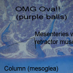
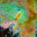
That right there is one gorgeous copepod, one of the bigger and more important groups of planktonic crustaceans. It looks huge but is actually tiny; probably 1-2mm. This is what they normally look like on a light microscope:

You can see how much richer and more detailed the top image is (although the colour is stained flouresence, not natural). It’s one of the winners in this years Nikon Small World competition, an annual contest for images taken with a microscope (photomicrographs). That particular image uses a technique called confocal microscopy, which uses lasers and clever optics to achieve great depth of field, i.e. where everything is in focus. NSW is close to my heart; I love photomicroscopy and even managed to scrape an honorable mention in the contest once. Meeting other microscopists at the award dinner I realised how utterly out my depth I was. The winner that year was this picture of a fly, which was composited from over a hundred images, using a computer algorithm to preserve the sharpest focus areas of each and layering them on top of each other… I havent entered since!
I bet you a beer you can’t resist wasting a chunk of time browsing the images from this and previous years winners; they’re just mesmerizing. As you do, consider my theory of winning NSW: it helps if your image looks like something else, like these crystals that look like a city skyline, or this one that looks like eyelashes, or this one that looks like a suspension bridge. There’s a whole world to photomicroscopy, and the techniques that go with it. Not all of these images were taken in the course of hard science; a lot of the time it’s a scientist who has noticed something that just looks cool, or they’re playing with some optical technique or other. Hard to argue with the results though: science as art.

>(although the colour is stained flouresence, not natural).
fluorescence!
Well yes, thanks so much for picking up on that
I can appreciate this. As a young rockhound (too long ago to mention) I used to mount microcrystal specimens under the microscope. Magnification up to 30X…anything higher
defeated the purpose. Never took photos. Now, old age eyesight and shaky fingers preclude any attempt, although I still have unmounted samples from my high school days. A good evenings work would result in maybe 3 specimens properly mounted.
Wow. This is very interesting. So it’s kind of like Macro but the micro of macro? This is amazing. You could actually make out the details of this teeny tiny organism. And yes I am scrolling through the 115 photos in the gallery and am currently mesmerized. I have got to show this to my friends. Thanks for sharing this ;)
HA! This made me almost spit my tea out with laughter – same problem for nematodes. They look SO CRAPPY when you look at them under light/dissection microscopes, severely hindering your attempts to convince undergrads and high schoolers of their ultimate awesomeness.